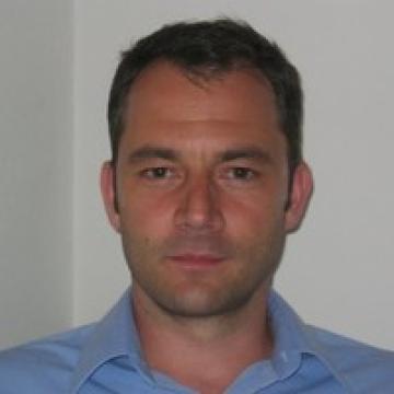
prof. Tom Van Hoof (PhD)

Principal investigator radiotherapy focused anatomy research unit
Full professor (Faculty of Medicine and Health Sciences, UGent)
Member of Biology department, evolutionary morphology unit (Faculty of Sciences)
Member of the Dutch Anatomy Society
Lecturer human anatomy for Physical Therapy and Rehabilitation Sciences
Research focus
My PhD focused on the 3D visualization and neurodynamics of the brachial plexus. My previously purely anatomical expertise rapidly became more clinically oriented when I started collaborating with the radiotherapy department. The brachial plexus is an organ at risk that needs to be spared from excessive radiation exposure during treatment. In order to spare the brachial plexus, it needs to be identified on the CT scans that are used for targeting the radiotherapy machinery. To avoid irradiation of the brachial plexus during radiation therapy, treatment guidelines were made during an anatomy and medical imaging oriented PhD that I helped supervise.
This new, patient oriented area of research was a great way to bring my anatomical research to the patient and the collaboration with the radiotherapy department continued with a second PhD project, this time focused on the lymphatic targets. The lymphatic targets, like the brachial plexus, are also extremely difficult to pinpoint on patient CT scans, so this second PhD focused on making a new set of guidelines for the prone crawl position that was recently developed by the radiotherapy department. Instead of sparing the lymphatics, like was the case with the brachial plexus, this time the lymphatics were the treatment target that needs to properly receive the prescribed dose, to prevent the cancer cells from spreading. This second PhD project unexpectedly opened up a whole new anatomical research area, because the lymphatics are on the verge of the visible and microscopic size range and they turned out to be wildly understudied. After successfully delivering the new set of guidelines for the lymphatic system, my research focus therefore switched to lymphatic micro-dissections and contrast agent injections in an effort to more accurately map the lymphatic system in 3D. To properly study the lymphatic system, a new research laboratory needed to be built within the anatomy department. This included the integration of a surgical microscope, a set up to perform lymphatic flow studies and a set up to assist with micro cannulations on a full body, making the anatomy department one of the few with a dedicated lymphatic anatomy research lab.
With my 20+ years of practical experience in the anatomy lab, I very much enjoy passing on my knowledge to the next generation of health care professionals through my lectures. At the same time, my collaborative research projects involving dissections, medical imaging and 3D medical imaging postprocessing also give me the opportunity to keep contributing to patient care.
Biography
Tom Van Hoof graduated for his master’s degree in Physiotherapy and Revalidation Sciences in 1997. The following two years he worked in the pharmaceutical industry as a product specialist (Fournier Pharma).
From 1999 until 2002 he studied Manual Therapy and he graduated for this specialisation in combination with managing his own physiotherapy practice dedicated to vertebral column rehabilitation and neurodynamics. At this time, neurodynamics was still a developing research field and being intrigued by the opportunity of contributing to this emerging field, he decided to trade the physiotherapy practice for an academic career.
In the spring of 2002 he started his own line of research ‘neurodynamics of the brachial plexus’ at the Anatomy Department of the Ghent University. As a predoctoral (mandate) assistant (2002-2008) he started teaching complete anatomy with specialities in the musculoskeletal and nervous system. During his time as predoctoral assistant he spent a great amount of time in the anatomy lab, teaching, researching and performing anatomical dissections. This love for and experience in the field of anatomy led to him being integrated in a managing role of the dissection lab, for the anatomy department. In this position, he spearheaded the integration of new embalming techniques for human specimens, which led to the introduction of a cadaver model that was suitable for surgical and endoscopic workshops (EndoGhent). He was also closely involved in the construction of a brand new dissection facility on the UZ Ghent campus. Once again being able to use his anatomical vision from a practical point of view, he was able to create what is to date one of the very few anatomy facilities that has reduced the use of the harmful embalming agent formaldehyde to a bare minimum, making one of the safest anatomy facilities in the world.
In 2008 he obtained his PhD in Medical Sciences for his research on 3D visualisation and neurodynamics of the brachial plexus. From this point forward, he became the point of contact for researchers from other, diverse disciplines in need of anatomical data. Broadening his research focus from the neck, shoulder and upper extremity meant developing a host of new research set ups in the anatomy lab and coming into contact with many different research methods from other fields. By integrating research techniques from the biomechanical field and by intensively being involved in imaging studies, many anatomical proof of concepts were developed as a step up to clinically applicable techniques.
Starting as a Tenure Track Professor in 2014 his previous experience in neurodynamic research and anatomical imaging studies led to a new research line within the field of radiotherapy. The focus of this research line was the development of anatomically verified radiotherapy guidelines for sparing the brachial plexus during radiation treatment. During his tenure track, he also endeavoured to create the Centre for Education, Training and Research in Anatomical Sciences, together with his anatomy department colleagues. For this purpose he used his anatomical knowledge to devise cadaver set ups that simulate surgical positions outside of the operating room. By using his expertise as a product specialist he built contacts within the medical industry, broadening the use of the human cadaver model from just research and teaching to an essential tool for training and product development for products like prosthesics and other implantable devices.
In 2019 Tom Van Hoof became Senior Lecturer and he officially became part of the Ghent University Radiotherapy department, as a dedicated anatomist. The brachial plexus project for the radiotherapy department evolved into a new anatomical research line to visualise the lymphatic system and to provide anatomically validated guidelines for radiotherapy of breast cancer patients. With these new guidelines being close to publication, the lymphatic system remains an intriguing target for more in depth anatomical study. For a dedicated anatomical researcher with nearly 20 years of experience in the anatomy lab, this highly understudied field provides the perfect opportunity to shine and develop new techniques that can be integrated in patient care.
In 2018 Tom Van Hoof obtained a Special Research Fund, Starting Grant to start his research to map the subclavian and axillary lymphatic system for the radiotherapy department.
In 2023 this project was followed by an FWO Junior postdoctoral grant for the continuation of the lymphatic research project of which Tom Van Hoof is the promotor.
Research team
- Prof. Dominique Adriaens (PhD) - full professor
- Prof. Carl Vangestel (PhD) - associate professor, biostatistician
- dr. Michael Stouthandel (PhD) - postdoctoral fellow
- dr. Joris Van de Velde (PhD) - education and research
Key publications
- The Lymphatic System in Breast Cancer: Anatomical and Molecular Approaches, Medicina, 2021, PMID 34833492
- Delineation guidelines for the lymphatic target volumes in 'prone crawl' radiotherapy treatment position for breast cancer patients, Scientific Reports, 2021, PMID 34795352
- Call for a Multidisciplinary Effort to Map the Lymphatic System with Advanced Medical Imaging Techniques: A Review of the Literature and Suggestions for Future Anatomical Research, The anatomical Record, 2019, PMID 31087787
- Optimal number of atlases and label fusion for automatic multi-atlas-based brachial plexus contouring in radiotherapy treatment planning, Radiation Oncology, 2016, PMID 26743131
- An anatomically validated brachial plexus contouring method for intensity modulated radiation therapy planning, International Journal of Radiation Oncology, Biology, Physics, 2013, PMID 24138919
- The effect of morphometric atlas selection on multi-atlas-based automatic brachial plexus segmentation, 2015, Radiation Oncology, PMID 26696278
- Morphometric atlas selection for automatic brachial plexus segmentation, International Journal of Radiation Oncology, Biology, Physics, 2015, PMID 25956831
- Using the venous angle as a pressure reservoir to retrogradely fill the subclavian lymphatic trunk with contrast agent for lymphatic mapping, Annals of Anatomy, 2020, PMID 32562859
- Application of frozen Thiel-embalmed specimens for radiotherapy guideline development: a method to create accurate MRI-enhanced CT datasets, Strahlentherapie und Onkologie, 2022, PMID 35403891
- The use of Thiel embalmed human cadavers for retrograde injection and visualization of the lymphatic system, The Anatomical Record, 2020, 31674142
Contact & links
- Lab address: K.L. Ledeganckstraat 35, Evolutionary morphology department, 3rd floor, room 37, Gent
- Google Scholar
- Professor Van Hoof and his team can provide assistance with anatomical research projects involving cadaver dissections and medical imaging on cadavers in the dissection facility
- Professor Van Hoof is interested to receive invitations for talks and presentations